
Digital X-Rays – Advanced Imaging for Accurate Diagnosis
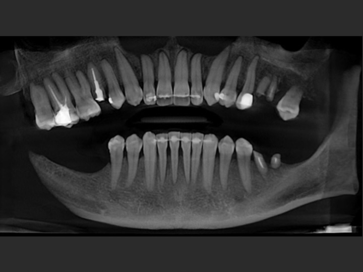
Digital radiography represents a modern advancement in dental imaging. Using highly sensitive digital sensors, digital X-rays reduce radiation exposure—often by 80–90% compared to traditional film—while providing clear, high-resolution diagnostic images. This enables precise evaluation while supporting patient safety and comfort.
In addition to full-size imaging systems, operatories are equipped with chairside digital X-ray units, allowing immediate, targeted images for focused diagnostic needs. These detailed images support efficient clinical decision-making and enhance patient comfort.
Uses of Digital X-Rays
Digital X-rays provide essential information not visible during a standard clinical exam, including:
-
Abscesses, cysts, or other infections
-
Bone loss from periodontal disease
-
Hidden decay between teeth
-
Tooth and root positioning
-
Developmental abnormalities
-
Conditions inside a tooth or below the gumline
-
Tumors, cysts, or other lesions
Early detection through digital imaging can support conservative treatment, reduce complexity, and help preserve natural teeth.
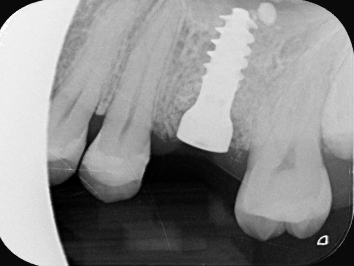
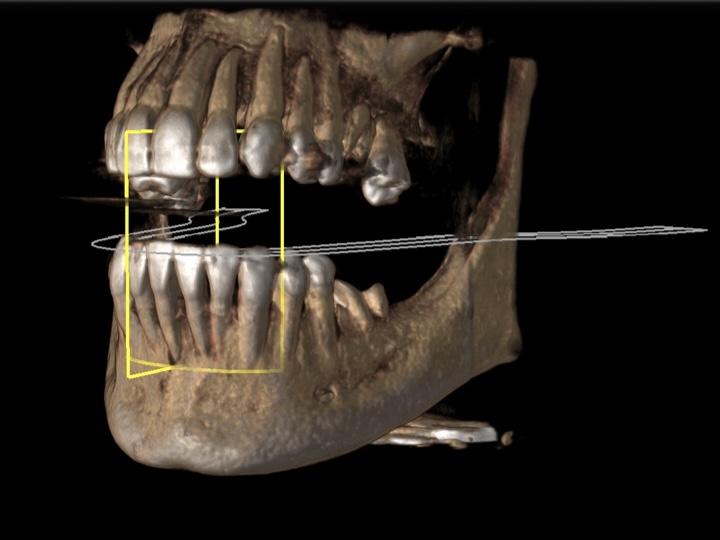
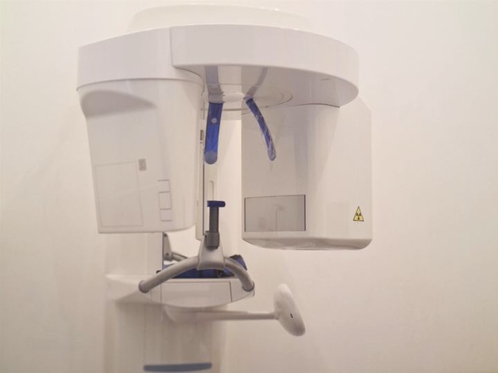
3D Imaging with CBCT Technology
For advanced diagnostics, an on-site CBCT (Cone Beam Computed Tomography) system provides detailed three-dimensional images of teeth, jawbone, joints, and surrounding structures. Unlike standard 2D radiographs, CBCT imaging reveals complex anatomical details with high precision.
CBCT imaging allows clinicians to:
-
Assess bone quality and need for bone augmentation
-
Plan dental implant placement with computer-guided precision
-
Analyze root canal anatomy for endodontic treatment
-
Evaluate impacted or ectopic teeth
-
Detect root resorption, developmental anomalies, and structural changes
-
Identify infections, bone lesions, or other pathologies
-
Examine trauma-related dental or bone injuries
-
Assess the paranasal sinuses and adjacent structures
CBCT is used selectively when enhanced anatomical detail is necessary for diagnosis or treatment planning.
Outcomes are individual and may vary according to each patient’s circumstances.
All images are the intellectual property of Dr. Catea and illustrate clinical work performed by him within the practice. Any reproduction, distribution, or use without prior written authorization is strictly prohibited.